Animal Tissues
In an animal,
individual cells differentiate during development to perform special functions
as aggregates called tissues.
A tissue (Fr. tissu, woven) is a group of similar cells
specialized for the performance of a common function. The study of tissues is
called histology (Gr. histos, tissue 1 logos,
discourse).
Animal tissues are classified as
1. 1. Epithelial tissue
2. 2. Connective tissue
3. 3. muscle tissue
4. 4. nervous tissue

1.
Epithelial Tissue:
Many Forms and Functions
Epithelial tissue
exists in many structural forms. In general, it
either covers or lines something and typically consists of renewable
sheets of cells that have surface specializations adapted for their specific
roles. Usually, a basement membrane separates epithelial tissues from
underlying, adjacent tissues. Epithelial tissues absorb (e.g., the lining of
the small intestine), transport (e.g., kidney tubules), excrete (e.g., sweat
and endocrine glands), protect (e.g., the skin), and contain nerve cells for
sensory reception (e.g., the taste buds in the tongue). The size, shape, and
arrangement of epithelial cells are directly related to these specific
functions.
Epithelial tissues
are classified on the basis of shape and the number of layers present. Epithelium
can be simple, consisting of only one layer
of cells, or stratified, consisting of two
or more stacked layers. Individual epithelial cells can be flat (squamous
epithelium;), cube shaped (cuboidal epithelium;), or column like (columnar
epithelium). The cells of pseudo-stratified ciliated
columnar epithelium possess cilia and appear stratified or layered, but they
are not, hence the prefix pseudo. They look layered because their nuclei are at
two or more levels within cells of the tissues and they grow in height as old
cells are replaced by new ones.
Types of Epithelium Tissue:

(a) Simple squamous
epithelium consists of a
single layer of tightly packed, flattened cells with a disk-shaped central
nucleus .
Location: Air sacs of the lungs, kidney glomeruli, lining of heart, blood
vessels, and lymphatic vessels.
Function: Allows passage of materials by
diffusion and filtration.
(b) Simple cuboidal epithelium consists of a single layer of tightly
packed, cube-shaped cells. Notice the cell layer indicated by the arrow.
Location: Kidney tubules, ducts and small glands, and
surface of ovary.
Function: Secretion and absorption.
(c) Simple columnar
epithelium consists of a
single layer of elongated cells. The arrow points to a specialized goblet cell
that secretes mucus.
Location: Lines digestive tract, gallbladder, and
excretory ducts of some glands.
Function:
Absorption, enzyme secretion.
(d) Pseudostratified ciliated columnar epithelium. A tuft of cilia tops each columnar cell,
except for goblet cells
Location: Lines bronchi, uterine tubes, and some regions of the uterus.
Function: Propels mucus or reproductive cells by ciliary action.
(e) Stratified
squamous epithelium consists of many layers of cells.

1.
Connective Tissue:
Connection and Support Connective tissues support and bind.
Unlike epithelial tissues, connective tissues are distributed
throughout an extracellular matrix. This matrix frequently contains fibers that
are embedded in a ground substance with a consistency anywhere from liquid to
solid. To a large extent, the nature of this extracellular material determines
the functional properties of the various connective tissues.
Connective tissues have two general types of fiber arrangement. In loose connective tissue, strong, flexible fibers of the protein collagen are interwoven with fine, elastic, and reticular fibers, giving loose connective tissue its elastic consistency and making it an excellent binding tissue (e.g., binding the skin to underlying muscle tissue). In fibrous connective tissue, the collagen fibers are densely packed and may lie parallel to one another, creating very strong cords, such as tendons (which connect muscles to bones or to other muscles) and ligaments (which connect bones to bones). Adipose tissue is a type of loose connective tissue that consists of large cells that store lipid. Most often, the cells accumulate in large numbers to form what is commonly called fat.

i.
Loose connective tissue contains numerous fibroblasts (arrows)
that produce collagenous and elastic fibers. Location: Widely distributed
under the epithelia of the human body. Function: Wraps and cushions organs.
ii.
Fibrous connective tissue consists largely of tightly packed
collagenous fibers.
Location: Dermis of the skin, sub-mucosa of the
digestive tract, and fibrous capsules of organs and joints.
Function: Provides structural strength.
iii.
Blood is a type of connective tissue. It consists of red blood cells, white
blood cells, and platelets suspended in an intercellular fluid (plasma) .
Location: Within blood vessels.
Function: Transports oxygen, carbon dioxide,
nutrients, wastes, hormones, minerals, vitamins, and other substances throughout
the bodies of animals.
(a) Adipose tissue cells (adipocytes) contain large fat droplets
that push the nuclei close to the plasma membrane. The arrow points to a
nucleus.
Location: Around kidneys, under skin, in bones, within abdomen, and in breasts. Function: Provides reserve fuel (lipids), insulates against heat loss, and supports and protects organs.
Cartilage
Cartilage is a hard
yet flexible tissue that supports structures such as the outer ear and forms
the entire skeleton of animals such as sharks and rays . Cells called
chondrocytes lie within spaces called lacunae that are surrounded by a rubbery
matrix that chondros-blasts secrete. This matrix, along with the collagen
and/or elastin fibers, gives cartilage its strength and elasticity. Bone cells
(osteocytes) also lie within lacunae, but the matrix around them is heavily
impregnated with calcium phosphate and calcium carbonate, making this kind of
tissue hard and ideally suited for its functions of support and protection.
a) Hyaline cartilage
cells are located in
lacunae (arrow) surrounded by intercellular material containing fine
collagenous fibers. Location: Forms embryonic skeleton; covers ends of
long bones; and forms cartilage of nose, trachea, and larynx. Function:
Support and reinforcement.
b) Elastic
cartilage contains fine collagenous fibers and many elastic fibers in its
intercellular material. Location: External ear, epiglottis. Function: Maintains
a structure’s shape while allowing great flexibility.
c) Fibrocartilage
contains many large, collagenous fibers in its intercellular material. The
arrow points to a fibroblast. Location: Intervertebral disks, pubic symphysis, and
disks of knee joint. Function: Absorbs compression shock.
d) Bone (osseous) tissue. Bone matrix is deposited in concentric layers around osteonic canals. Location: Bones. Function: Supports, protects, provides lever system for muscles to act on, stores calcium and fat, and forms blood cells.
3. Muscle Tissue:
Movement Muscle tissue allows movement. The three kinds of muscle tissue are skeletal, smooth, and cardiac. Skeletal muscle is attached to bones and makes body movement possible in vertebrates. The rhythmic contractions of smooth muscle create a churning action (as in the stomach), help propel material through a tubular structure (as in the intestines), and control size changes in hollow organs such as the urinary bladder and uterus. The contractions of cardiac muscle result in the heart beating.
(a) Skeletal muscle
tissue is composed of
striated muscle fibers (cells) that are long and cylindrical and contain many
peripheral nuclei. Location: In skeletal muscles attached to bones. Function:
Voluntary movement, locomotion.
(b) Smooth muscle tissue is formed of spindle-shaped cells, each
containing a single centrally located nucleus (arrow). Cells are arranged
closely to form sheets. Smooth muscle tissue is not striated. Location: Mostly
in the walls of hollow organs. Function: Moves substances or objects
(foodstuffs, urine, a baby) along internal passageways; involuntary control.
(c) Cardiac muscle tissue consists of branched striated cells, each containing a single nucleus and specialized cell junctions called intercalated disks (arrow) that allow ions (action potentials) to move quickly from cell to cell. Location: The walls of the heart. Function: As the walls of the heart contract, cardiac muscle tissue propels blood into the circulation; involuntary control.
4.
Nervous Tissue:
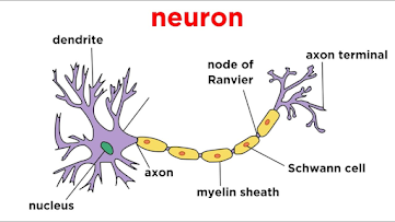
Communication Nervous tissue is composed of several different types of cells: Impulse-conducting cells are called neurons. cells involved with protection, support, and nourishment arecalled neuroglia; and cells that form sheaths and help protect, nourish, and maintain cells of the peripheral nervous system are called peripheral glial cells.
Neurons in nervous
tissue transmit electrical signals to other neurons, muscles, or glands. Location: Brain,
spinal cord, and nerves. Function: Transmits electrical signals from sensory
receptors to the spinal cord or brain, and from the spinal cord or brain to
effectors (muscles and glands).


Post a Comment